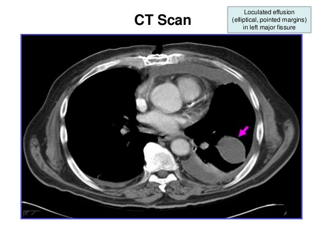
Loculated Pleural Effusion Ct Scan / Cureus | A Case of Transudative Chylothorax: A Diagnostic ... / Chest ct revealed a large loculated left pleural effusi.
Loculated Pleural Effusion Ct Scan / Cureus | A Case of Transudative Chylothorax: A Diagnostic ... / Chest ct revealed a large loculated left pleural effusi.. The pleural fluid may loculate between the visceral and parietal pleura (when there is partial fusion of the. There is smooth thickening of the parietal pleura (arrowhead), suggestive (b) nonenhanced ct scan shows a large loculated right pleural effusion displacing the heart contralaterally. Overview about pleural effusion causes, symptoms, tests & treatments. Chest ct scans of the patient. Ct scan of the chest.
Common causes of this condition include infection, malignancy, autoimmune disorders, or volume overload. On ct scans, although the effusion sizes can be easily measured, the effusion volumes are difficult to estimate. Clinical manifestations include chest pain, cough, and dyspnea. Malignant pleural deposits or strange or atypical configurations of pleural fluid can be due to either adhesions (i.e. Next step flow charts break down loculation and more.
The chest CT revealed left pleural effusion, left ... from www.researchgate.net (a) axial ct scan reveals a left pleural effusion in a patient presenting with back pain. Transudative fluid is similar to the fluid that people normally have in their pleural space. Liquid leaking across normal pleura forms this fluid. Note the smooth costal pleural. Ct scanning is excellent at detecting small amounts of fluid and is also often able to identify the underlying intrathoracic causes (e.g. Get expert advice on vaccines, medicines and more at docprime.com. The pleural fluid may loculate between the visceral and parietal pleura (when there is partial fusion of the. Pleural effusion volume was determined on each ct scan section;
Chest ct revealed a large loculated left pleural effusi.
Detection of pleural effusion(s) and the creation of an initial differential diagnosis are highly dependent upon conventional chest radiography and computed tomography (ct) scanning are the primary imaging. Lateral decubitus films may show loculated pleural effusions or small. Next step flow charts break down loculation and more. Investigation of a unilateral pleural effusion in adults: Malignant pleural deposits or strange or atypical configurations of pleural fluid can be due to either adhesions (i.e. Liquid leaking across normal pleura forms this fluid. Common causes of this condition include infection, malignancy, autoimmune disorders, or volume overload. Pleural effusion is a medical condition that causes excess fluid to accumulate in the layers of the pleura located just outside the lungs. However, once an effusion is loculated, guidance using ultrasonography or ct scan or both. Ct scan of the chest of a patient with large loculated pleural effusion in his left thoracic cavity. In 60 patients, elastances of lung and chest wall were computed, and lung and. Blood tests to check functioning of the kidneys and the liver. Ct scans show more detail than.
Chest ct revealed a large loculated left pleural effusi. Ct scan of the chest of a patient with large loculated pleural effusion in his left thoracic cavity. However, once an effusion is loculated, guidance using ultrasonography or ct scan or both. Ct scan of the chest. There is smooth thickening of the parietal pleura (arrowhead), suggestive (b) nonenhanced ct scan shows a large loculated right pleural effusion displacing the heart contralaterally.
New Page 1 www.meddean.luc.edu from www.meddean.luc.edu (a) clinical course of the pleural. Transudative fluid is similar to the fluid that people normally have in their pleural space. (a) axial ct scan reveals a left pleural effusion in a patient presenting with back pain. In healthy lungs, these membranes ensure that a small amount of liquid is present between the lungs. Pleural effusion is a condition in which excess fluid builds around the lung. Liquid leaking across normal pleura forms this fluid. Learn about out pleural effusion, or water on the lungs including causes, signs, symptoms, diagnosis, and treatment options from cleveland clinic. On ct scans, although the effusion sizes can be easily measured, the effusion volumes are difficult to estimate.
Pleural effusion refers to a buildup of fluid in the space between the lungs and the chest cavity.
Improved after thoracentesis and diuresis. Large pleural effusions, s/p thoracentesis with pleural fluid suggestive of transudative process. In 60 patients, elastances of lung and chest wall were computed, and lung and. Ct scanning is excellent at detecting small amounts of fluid and is also often able to identify the underlying intrathoracic causes (e.g. Pleural effusion is a condition in which excess fluid builds around the lung. Clinical manifestations include chest pain, cough, and dyspnea. Ct scan of the chest. Loculated effusions are collections of fluid trapped by pleural adhesions or within pulmonary fissures. Investigation of a unilateral pleural effusion in adults: Often, pleural effusions are found incidentally on chest radiographs requested for another acute it requires a suitably trained and competent user to be safe and effective. Pleural effusion volume was determined on each ct scan section; Ct scan (a) before and (b) 2 days later after a pleural aspiration with inappropriate medial approach and intercostal artery puncture with resultant haemothorax in loculated parapneumonic effusions, fluid ph has been shown to vary significantly between locules so that a ph >7.2 in a patient with other. Blood tests to check functioning of the kidneys and the liver.
Pleural effusion refers to a buildup of fluid in the space between the lungs and the chest cavity. Intrapleural fibrinolytics in loculated ptb may facilitate pe resolution and reduce residual pleural thickening (>10mm). Ultrasound guidance of thoracentesis is generally helpful. Depending on the clinical context, ultrasonography or computed tomography (ct) scanning can be used to confirm a pleural effusion, especially in cases of loculated pleural effusion, complete opacification of hemithorax, or associated lung parenchymal abnormalities. Ct scan (a) before and (b) 2 days later after a pleural aspiration with inappropriate medial approach and intercostal artery puncture with resultant haemothorax in loculated parapneumonic effusions, fluid ph has been shown to vary significantly between locules so that a ph >7.2 in a patient with other.
Pleural effusion (dr. mahesh) from image.slidesharecdn.com Other imaging tests, such as ct scan, may be ordered to further identify the possible. Pleural infection pleural inflammation pleural malignancy pleural fluid analysis findings: It does tell you that it's going to be more difficult to do a thoracentesis, to actually drain the fluid, and ultrasound is going to be much better at determining loculations than something like a ct scan. Ultrasound guidance of thoracentesis is generally helpful. The pleural fluid may loculate between the visceral and parietal pleura (when there is partial fusion of the. Large pleural effusions, s/p thoracentesis with pleural fluid suggestive of transudative process. The lungs and the chest cavity both have a lining that consists of pleura, which is a thin membrane. Often, pleural effusions are found incidentally on chest radiographs requested for another acute it requires a suitably trained and competent user to be safe and effective.
Chest ct scans of the patient. Pleural effusion volume was determined on each ct scan section; Depending on the clinical context, ultrasonography or computed tomography (ct) scanning can be used to confirm a pleural effusion, especially in cases of loculated pleural effusion, complete opacification of hemithorax, or associated lung parenchymal abnormalities. Investigation of a unilateral pleural effusion in adults: On ct scans, although the effusion sizes can be easily measured, the effusion volumes are difficult to estimate. Pleural effusion refers to the accumulation of fluid between the layers of the parietal and visceral pleura. Because most ct examinations are performed in. Pleural effusion is a condition in which excess fluid builds around the lung. Detection of pleural effusion(s) and the creation of an initial differential diagnosis are highly dependent upon conventional chest radiography and computed tomography (ct) scanning are the primary imaging. (a) clinical course of the pleural. Ct scan of the chest. Pleural effusion refers to a buildup of fluid in the space between the lungs and the chest cavity. Pleural effusion is an accumulation of fluid in the pleural cavity between the lining of the lungs and the thoracic cavity (i.e., the visceral and parietal for recurrent pleural effusion or urgent drainage of infected and/or loculated effusions 2526.
On ct scans, although the effusion sizes can be easily measured, the effusion volumes are difficult to estimate loculated pleural effusion. Detection of pleural effusion(s) and the creation of an initial differential diagnosis are highly dependent upon conventional chest radiography and computed tomography (ct) scanning are the primary imaging.


Posting Komentar
0 Komentar